The inverted microscope has the characteristics of microscopic observation in a culture bottle or petri dish, and can observe transparent living bodies without staining; epi-fluorescence microscopy is suitable for fluorescence microscopy. The instrument is especially suitable for microscopic research on living cells and tissues, fluids, sediments, etc. It is an ideal instrument for research work in biology, cytology, oncology, genetics, immunology and so on. It can be used by scientific research, universities, medical treatment, epidemic prevention, agriculture and animal husbandry and other departments. The inverted fluorescence microscope is composed of an inverted microscope and an epi-fluorescence microscope. The instrument is equipped with long working distance plan achromatic objectives, large field eyepieces, and binocular observation. The inverted microscope is also equipped with extra long or super long working distance condensers. It is also equipped with phase contrast devices and long working distance flat field phase contrast objectives.
Inverted fluorescence microscope is a new type of fluorescence microscope developed in modern times. It is characterized by the excitation light falling from the objective lens to the surface of the specimen. That is, the same objective lens is used as the illumination condenser and the objective lens for collecting fluorescence. A two-color beam splitter needs to be added to the optical path, which is 45 degrees to the optical uranium. At the angle, the excitation light is reflected into the objective lens and concentrated on the sample. The fluorescence generated by the sample and the excitation light reflected by the objective lens surface and the cover glass surface enter the objective lens at the same time, and return to the two-color beam splitter to make the excitation light Separated from fluorescence, the residual excitation light is then absorbed by the blocking filter. For example, the combination of different excitation filter / dual-color beam separator / blocking filter can meet the needs of different fluorescent reaction products. The advantage of this type of fluorescence microscope is that the illumination of the field of view is uniform, the imaging is clear, and the greater the magnification, the stronger the fluorescence.
The inverted fluorescence microscope is composed of a fluorescent accessory and an inverted microscope. It is mainly used for fluorescence and phase contrast observation of living tissues such as cells. The inverted microscope (Inverted microscope) is to adapt to microscopic observations in tissue culture, cell culture in vitro, plankton, environmental protection, food inspection, etc. in the fields of biology and medicine. Since these living objects are placed in a Petri dish (or culture bottle), the working distance between the objective lens and condenser lens of the inverted microscope is required to be able to directly observe and study the detected objects in the Petri dish. Therefore, the positions of the objective lens, condenser lens and light source are reversed, so it is called "inverted microscope". Inverted microscopes are mostly used for colorless and transparent living observation. Add a set of fluorescent accessories on the basis of inverted microscopes: laser excitation block, fluorescent light source, fluorescent illuminator, excitation block switching device to perform inverted fluorescence observation.
The imported microscopes of the four major brands all have corresponding inverted fluorescence microscopes, such as Olympus' IX71 inverted fluorescence microscope and CKX41 inverted fluorescence microscope. In recent years, with the continuous improvement of phase contrast technology and fluorescence imaging technology, domestic brand microscope manufacturers have also introduced a variety of inverted fluorescence microscopes. The Sino-foreign joint venture Aopu Optoelectronics DSY5000X inverted fluorescence microscope is one, which has high fluorescence brightness and clear imaging. Or close to the level of similar mid-range microscopes abroad.
DSY5000X inverted biological microscope
1. Optical system: UCIS optical system with independent correction of infinity chromatic aberration
2. Frame: integrated structure, ergonomic principles, stable and reliable, national appearance patent
3. Observation tube: binocular observation tube, tilt angle 45 °, pupil distance adjustment 52-75mm, adjustable diopter
4. Eyepiece: WF10X / 20mm flat field wide field eyepiece, high eye point, pupil observation distance 21mm
5. Objective lens:
10X Infinity long distance plan achromatic objective NA0.25 WD9.67
20X Infinity long-range plan achromatic objective NA0.40 WD7.97
40X Infinity long distance plan achromatic objective NA0.60 WD3.76
20X Infinity long distance plan field positive phase contrast achromatic objective lens NA0.40 WD7.97
6. Stage: fixed stage 240mmX260mm; equipped with low coaxial flexible XY movement adjustment handwheel, moving range 135mmX85mm, equipped with water drop carrier (Φ118), multifunctional carrier (76 X 26, Φ60) .
7. Objective lens converter: Five-hole positioning converter, ball bearing positioning, with anti-mildew device.
8. Coarse and fine adjustment: Coarse and fine adjustment coaxial, equipped with limit device and locking device, low hand position coaxial focusing hand wheel, fine adjustment hand wheel grid value 0.001mm, focusing is more accurate.
9. Condenser lens: Super long working distance condenser lens 72mm, numerical aperture N.A0.30, equipped with three-hole phase contrast ring plate, equipped with three color filter sets of yellow, green and blue.
10. Light source: built-in aspheric lens, 6V30WOsram halogen lamp, wide voltage 110-240V, 50 / 60Hz, equipped with UOP special power box.
11. Color filter: blue filter group, green filter group, yellow filter group.
12. C interface: 0.5XC interface
13. ISO9001, ISO14001 certification, Chongqing key new product certificate.
14. Sino-foreign joint ventures, national high-tech enterprises.
Fluorescence accessories: Epi-fluorescence device, fluorescent light box, fluorescent power box, B, G, UV, V fluorescent excitation filter
gain
40X ~ 960X
eyepiece
Flat field 10x high eyepoint eyepiece 20mm field of view, pupil distance 21mm, diopter adjustable
Objective lens
Infinity long-distance plan objectives 4X (optional), 10X, 20X, 40X, 60X (optional)
Infinite long distance flat field phase contrast 10X, 20X, 40X
Observation tube
Hinged binocular, tilted at 45 °, interpupillary distance 52mm ~ 75 mm, adjustable diopter
Hinged trinocular, 45 ° tilt, interpupillary distance 52mm ~ 75 mm, adjustable diopter
Objective lens converter
Internal positioning five-hole converter
Coarse and fine focusing device
Low-level coarse and fine coaxial focusing handwheel; micro-moving handwheel 0.1mm / rev, grid value 0.001mm; coarse-motion tightness adjustable; upper limit device of working table.
Stage
Fixed stage 240mmX260mm; equipped with low coaxial flexible XY movement adjustment handwheel, movement range 135mmX85mm
Condenser
NA0.70 WD120mm
Phase contrast ring plate
10X, 20X, 40X
C interface
0.5X, 1X
1. Requirements for the preparation of fluorescent microscope specimens
1. Slides
The thickness of the slide glass should be between 0.8 and 1.2 mm. A too thick slope film, on the one hand, absorbs much light, and on the other hand, it cannot concentrate the excitation light on the specimen. The slides must be smooth, uniform in thickness, and have no obvious autofluorescence. Sometimes quartz glass slides are required.
2. Cover glass
The thickness of the coverslip is about 0.17mm, smooth and clean. In order to enhance the excitation light, dry coverslips can also be used. This is a special cover glass coated with several layers of substances (such as magnesium fluoride) that interfere with different wavelengths of light. Passing, while reflecting the excitation light, this reflected excitation light can excite the specimen.
3. Specimens
Tissue slices or other specimens should not be too thick. For example, if the excitation light is too thick, most of the excitation light is consumed in the lower part of the specimen, and the upper part directly observed by the objective lens cannot be fully excited. In addition, cells overlap or impurities cover up, affecting judgment.
4. Mounting agent
The glycerin commonly used as the mounting agent must be free of spontaneous fluorescence, colorless and transparent. The brightness of the fluorescence is brighter at pH 8.5 to 9.5, and it is not easy to fade quickly. Therefore, an equal amount of mixed solution of glycerin and carbonate buffer of 0.5mol / L PH9.0 ~ 9.5 is commonly used as a mounting agent.
5. Mirror oil
Generally, when using dark field fluorescence microscope and oil lens to observe the specimen, lens oil must be used. It is best to use a special non-fluorescent lens oil, which can also be replaced by the above glycerin, and liquid paraffin can also be used, but the refractive index is low, which has a slight impact on image quality.
2. Basic operation of fluorescence microscope
1. Turn off the electric light in the room and turn on the mercury lamp of the microscope;
2. Select the corresponding filter according to the sample labeled fluorescein;
3. Place the sample and find a suitable field of view;
4. If you need to take pictures, please make sure that the color film is installed in the camera (it is best to use 27 fixed film);
5. Turn on the self-timer and select the manual mode, usually the shooting speed is within 0.5-10 seconds;
6. At the end of use, turn off all power and make a record of use.
Third, the recording method of fluorescent images
The fluorescence images seen by a fluorescence microscope have morphological characteristics and fluorescence color and brightness. When judging the results, the two must be combined to make a comprehensive judgment. The results are recorded based on subjective indicators, that is, the worker's eyesight. As a general qualitative observation, it is basically reliable. With the development of technical science, objective indicators are used to record the judgment results in different degrees, such as cell spectrophotometer, image analyzer and other instruments. But the results recorded by these instruments must also be combined with subjective judgment.
Fluorescence microscopy photography technology is necessary to record fluorescence images. Since fluorescence is easily faded and weakened, the results should be photographed immediately. The method is basically the same as that of ordinary photomicrography. Just need to use high-speed photosensitive film. Due to the great effect of ultraviolet light on the fluorescence quenching, such as FITC markers, the fluorescence brightness is reduced by 50% when irradiated with ultraviolet light for 30s. Therefore, if the exposure speed is too slow, the fluorescent image cannot be taken. General research-type fluorescence microscopes have semi-automatic or fully automatic photomicrography system devices.
4. Precautions for using fluorescence microscope
1. Operate strictly in accordance with the requirements of the fluorescence microscope factory manual, and do not change the program at will.
2. Should be checked in a dark room. After entering the dark room, connect the power supply and ignite the ultra-high pressure mercury lamp for 5 to 15 minutes. After the light source emits strong light to stabilize, the eyes are fully adapted to the dark room, and then start observing the specimen.
3. To prevent damage to eyes caused by ultraviolet rays, wear protective glasses when adjusting the light source.
4. The inspection time is preferably 1 ~ 2h each time, more than 90min, the luminous intensity of the ultra-high pressure mercury lamp gradually decreases, and the fluorescence weakens; after the specimen is exposed to ultraviolet light for 3 ~ 5min, the fluorescence also significantly decreases; .
5. The life of fluorescent microscope light source is limited. Specimens should be inspected intensively to save time and protect the light source. When it is hot, the fan should be added to cool down, and the new lamp should be recorded from the beginning. When the lamp is extinguished and you want to use it again, you can only ignite it after the bulb has cooled down sufficiently. Avoid lighting the light source several times a day.
6. Observe the specimen immediately after staining, and the fluorescence will gradually weaken over time. If the specimen is stored in a polyethylene plastic bag at 4 ° C, it can delay the fluorescence attenuation time and prevent the mounting agent from evaporating.
7. Judgment standard of fluorescence brightness: generally divided into four levels, namely "-" means no or visible weak fluorescence. "+" Means that only clearly visible fluorescence can be seen. "++" means that bright fluorescence is visible. "+++" refers to visible dazzling fluorescence.
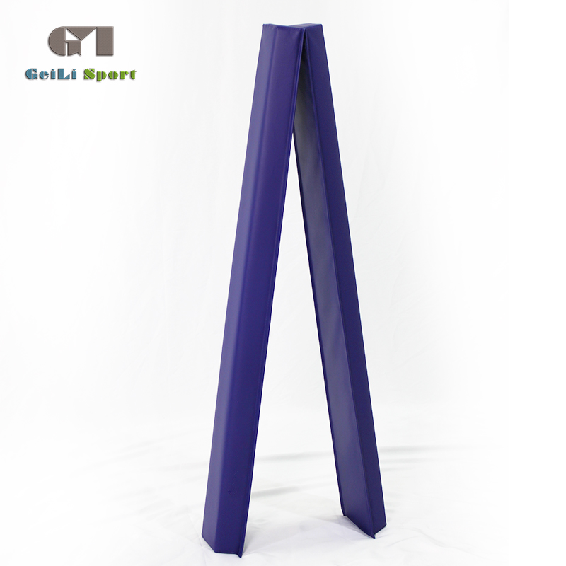
Now you do not have to worry about your enthusiastic kid who loves gymnastics more than anything but fails to practice just as much because of the lack of opportunities. The gymnastics balance beams are here to solve the problem as it can be set-up in your own house and your kid can continue polishing her moves as much he/she wants to without any time limits. For better performances in competitions, most of these beams are made out of materials that are similar to the ones used in competitions, thus no hassles of adjusting to a different beam. Moreover, they ensure quality and stability to avoid any risks of damages that can occur due to low-quality balance beams.we have many different balance beams,various colors and sizes.


Foam Balance Beam,Outdoor Balance Beam,Adjustable Balance Beam,Gymnastics Balance Beam
Shaoxing geili sports and leisure goods Co.,Ltd , https://www.geilisports.com