When it comes to biology, everyone will think of a different kind of cell. But to observe the subtle changes in cell morphology, a cost-effective Microscope is essential. Since Levin Hook invented the first optical microscope, the microscope has been constantly updated to meet the needs of the observer. At present, the most commonly used biological cell culture is a fluorescence microscope, which can be used to observe three kinds of fluorescence of green, red and blue. In addition, from the perspective of living cells and fixed cells, it can be divided into inverted microscope and upright microscope.
Let me talk about the fluorescence microscope of Olympus. The machine model used by Kamaishu is OLYMPUS BX51 (picture from the network).

The reason why many people choose Olympus's fluorescence microscope is largely because their software design is quite human. This software is very powerful, from image capture to image post-processing in one step, to meet a variety of shooting details you can think of and can not think of. In terms of image acquisition, the software's live image capture pixels are relatively high, and the sharpness is also very good. In addition, an image lock button is also provided, which is convenient for locking the observation area and facilitating the collection of multiple photos. When preparing to shoot, first make a white balance of the photo, which is very important for any shooting. Olympus's fluorescence microscope is designed with two white balance adjustment modes. In the white light mode, with a square focus frame of about 1 cm as the center, select one touch or peripheral block selection mode. It is easy to complete the white balance setting. In fluorescence mode, the focus frame needs to be shrunk to the fluorescent area before white balance correction can be performed. In addition, his family's lens is also quite clear, also equipped with a live map shooting mode, support for a period of animation shooting, as long as the shooting time range, with a culture box can achieve dynamic shooting of live cells. In terms of image processing, the function is also relatively complete, you can add a ruler, you can also achieve the overlapping effect of multi-color pictures, and the interface is very friendly, photographers who are not familiar with photoshop, if you have this software, you will feel convenient a lot of. It is said that Olympus's fluorescence microscope software sold very well in the past few years, but in recent years it has been affected by piracy and sales have fallen rapidly. After all, this set of tens of thousands of software, in addition to the powerful, the price is quite expensive, many sales agents Olympus fluorescent microscope companies in order to reduce costs, often to install pirated software for customers, it has affected The amount of software sold. Having said so many advantages, what is the disadvantage of Olympus's fluorescence microscope? I think it is more than 40 times the resolution of the image, because the clarity of the observation will be affected by the objective lens more than 20 times. Although his home also provides a lens switching mode, the improvement is not obvious. However, if you don't mind trying oil mirrors, Olympus's fluorescence microscope provides a 100x objective lens, and the results after the oil is dripping can be satisfactory, even the small organelles in the cells and the fluorescence produced by transfection. Small particles can sometimes be observed very clearly and can provide good evidence for the publication of the article.
The other thing to say is the recent Leica fluorescence microscope, modeled Leica DMI 300B (picture from the network).
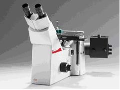
To be honest, Kamaishu only learned about this German brand in recent use, and the appearance of the product feels good. According to their engineers, their lens price is relatively high. In use, I also think that the lens clarity of his home can be regarded as the leader in low-priced products. And for those labs that don't have enough funds, buy an inverted microscope, or ask the engineer to do a small modification at the same time, it can be used as a positive and negative dual-use machine, and it is also a good choice. But there is a fatal weakness, that is, his family's software, super bad. Although there are two interfaces in Chinese and English, there are many disadvantages. Firstly, the adjustment of white balance is rough. In addition, the animation of dynamic animation is not supported, and the capture pixels of the live image are too low, which often causes the picture to appear sluggish and the focal length is not accurate. The phenomenon. In the subsequent processing of the image, it also has very little function. It must be helped by other software such as photoshop, so I can only say that the Leica fluorescence microscope is a device that is quite mismatched between hardware and software. I personally do not recommend his software. If you want to install an Olympus software, the engineers at his home are charged a lot, and the individual feels a bit pit.
The eye is the window of the soul, and the microscope is the soul carriage of the biologist, which can help us lead to more mysterious and unknown biological fields. We must not only know how to choose, but also how to control this carriage in order to achieve twice the result with half the effort. A good lens plus good software is the two powerful reins, which are indispensable. How to DIY a cost-effective microscopic observation equipment, my suggestion is to shop around, consider comprehensively, from cost to benefit, and then make a decision that is in line with its own positioning.
This life-size digestive system model demonstrates the entire digestive system in graphic relief. It features: nose, mouth cavity and pharynx, esophagus, GI
tract, liver with gallbladder, pancreas and spleen. The duodenum, caecum and rectum of the digestive system are opened. The products are made of imported PVC materials, environmental protection paint, computer color matching and artificial color painting. Every product has been strictly controlled before entering the market. All models were taken in kind.
Digestive Model,Human Digestive Model,Digestive System Model,Digestive Anatomy Model
Xinxiang Vic Science&Education Co.,Ltd. , https://www.hnhumananatomymodel.com